About this service
An ultrasound examination obtains images of the body without the use of x-rays.
It uses high frequency sound waves which pass through the skin and are reflected by the body’s internal organs. These ‘echoes’ are converted, with the aid of a computer, into a picture onto a screen.
We perform a variety of ultrasound scans across Warrington Hospital, the Nightingale building, the Captain Sir Tom Moore building and Halton Health Hub.
Opening times:
Warrington Hospital: 8am-8pm Monday to Thursday; 8am-6pm Friday.
Halton Nightingale: 8.30am–5pm Monday to Friday.
Captain Sir Tom Moore building: 8.30am–5pm Monday to Friday.
Halton Health Hub: 8.30am–4.30pm Monday to Friday.
Weekend sessions are sometimes available.
In addition to our main ultrasound scan departments, we also have ultrasound machines located across the Trust within the early pregnancy assessment unit, antenatal day unit, antenatal clinic, vascular lab and rapid access clinics.
Some of our areas have self check in. If not, a member of the administration and clerical team will greet you when you arrive at one of our receptions. They will check that we have all your details and that they are correct before booking you in. Our admin team includes secretaries, booking staff who will book appointments and answer queries, supervisors, and managers.
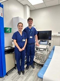
Ultrasound scans can be performed by doctors, sonographers, midwife sonographers, or vascular scientists – All of which are highly qualified members of staff who report their own examinations.
Radiology Department Assistants (RDAs) support the Ultrasound team to help and care for patients. They will help prepare patients for examination, insert cannulas where required and act as chaperones.
Porters are incredibly important, moving patients on beds, trolleys, or in wheelchairs between wards, departments, and Radiology.
For all General Ultrasound enquiries, please telephone: 01925 275588
For all Obstetric (pregnancy) Ultrasound enquiries, please telephone: 01925 662311
Where are we?
| Ultrasound Radiology Dept Appleton Wing Warrington Hospital Warrington Cheshire WA5 1QG |
Ultrasound Radiology Department Captain Sir Tom Moore building Earls Way Palacefields Runcorn WA7 2HH |
Ultrasound Nightingale building Hospital Way, Palacefields, Runcorn WA7 2DA |
Ultrasound Halton Health Hub Runcorn Shopping City, Palacefields, Runcorn WA7 2EU |
Limitations of Ultrasound
Ultrasound waves are disrupted by air or gas; therefore, ultrasound is not an ideal imaging technique for air-filled bowel or organs obscured by the bowel. Patients who are overweight or obese are more difficult to image by ultrasound because greater amounts of tissue weaken the sound waves as they pass deeper into the body. This results in images that are blurry and more difficult to see some pathology. Ultrasound also has difficulty penetrating bone and, therefore, can only see the outer surface of bony structures and not what lies within (except in babies who have more cartilage in their skeletons than older children or adults).
Mobile phone policy
We understand that often a family’s visit to the ultrasound department can be an exciting time. However, patients are respectfully reminded that all scans performed in the ultrasound department are medical examinations and therefore the use of mobile phones or other recording equipment is not permitted.
Ultrasound results
Finding out the results from your ultrasound examination depends upon the type of ultrasound scan you are having. Sometimes the results of the examination may be discussed immediately or sometimes the results will be made available in 1 -2 weeks time after being delivered to the health care professional that requested the ultrasound. Sometimes ultrasound findings require further clarification, and you may require an additional form of imaging such as an X-ray, CT scan or MRI scan in order to get a result.


